
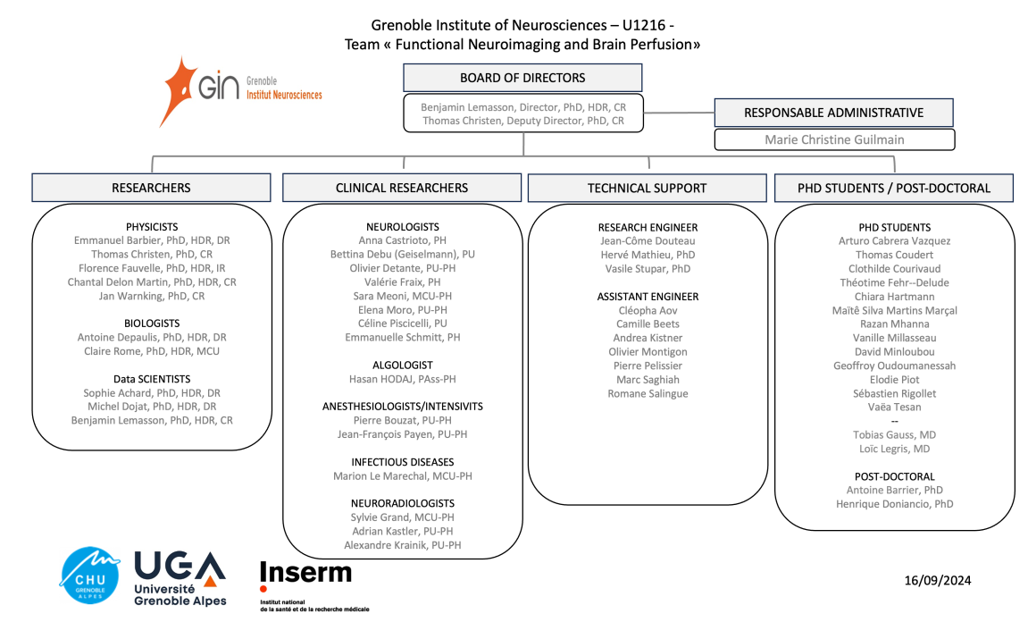
Dirigée par Benjamin LEMASSON et Thomas CHRISTEN
Thèmes de recherche
L'équipe est à l’origine de méthodes reconnues comme innovantes pour le traitement et l’analyse de données en imagerie et spectroscopie par résonance magnétique nucléaire (IRM et SRM) du fait de la complémentarité entre physiciens, mathématiciens et spécialistes du vivant (cliniciens, biologistes notamment) travaillant conjointement.
Innover et développer des méthodes d’acquisition et d’analyse d’images répondant aux besoins actuels de la recherche clinique et préclinique nécessite d’identifier les applications clés et d’effectuer leur évaluation rigoureuse. Afin d’améliorer la quantité et la qualité des informations obtenues par imagerie et spectroscopie par résonance magnétique nucléaire (IRM et SRM), l’équipe « Neuroimagerie fonctionnelle et perfusion cérébrale » développe les projets de recherches suivants :
- Des méthodes d’acquisition en IRM physiologique et fonctionnelle plus précises : Grâce au développement de méthodes originales pour caractériser la vasoréactivité et l’oxygénation cérébrale, à la maitrise de l’ensemble des outils de caractérisation de la perfusion cérébrale, et à l’aide d’outils d’analyses originaux (collab. Inria, Neurospin), nous souhaitons développer une application robuste de l’IRM fonctionnelle (IRMf) et de l’imagerie de la connectivité fonctionnelle en préclinique et en clinique, notamment dans des situations où le couplage entre capillaires et neurones est altéré.
- Identifier des biomarqueurs d’imagerie innovants : améliorer l’exploitation des nombreuses informations accessibles par IRM et SRM en développant des outils automatiques d’extraction, de quantification et de positionnement de ces données dans des atlas de référence pour être ensuite transmises au clinicien. Le logiciel « LOCUS » localisant en quelques minutes les tissus et structures du cerveau malgré la qualité variable des images obtenues par IRM est un exemple de méthode innovante développée récemment. Ce logiciel a été étendu pour segmenter automatiquement des lésions pour la sclérose en plaques, l’accident vasculaire cérébral, les tumeurs cérébrales, le traumatisme crânien, l'épilepsie où la maladie de Parkinson. Nous mettons également en oeuvre des outils de classifications (statistiques avancée, intelligence artificielle).
- Guider la thérapie par imagerie : mettre au point des protocoles multi-thérapeutiques complexes en utilisant les performances de l’imagerie en évaluant l’effet de chaque drogue utilisée et les éventuelles synergies entre elles, en renforçant l’emploi de certaines drogues et en guidant la libération localisée de drogues, ce qui permet également de réduire les effets secondaires.
Techniques utilisées
Acquisition de données
- L'imagerie et la spectroscopie par résonance magnétique nucléaire (IRM et SRM), en lien étroit avec la plateforme IRM de Grenoble (UMS IRMaGE), une composante de la plateforme d'imagerie in vivo de Grenoble, (labelisée IBiSA). L'équipe et la plateforme IRM de Grenoble sont partenaires de l'infrastructure nationale en imagerie du vivant, FLI (Grand Emprunt 2012).
- L'histologie et l'immuno-histochimie
- Simulations numériques de phénomènes physiques et biophysiques (approches in silico)
Analyse d'images
-
Modèles markoviens distribués pour la segmentation d'IRM cérébrale (logiciel Locus et P-LOCUS)
-
Cartographie rétinotopique fonctionnelle (logiciel Brain_A_La_Carte), rétinotopie inverse (logiciel RIME), Analyse d'images multiparamétriques (logiciel MP3)
-
Logiciels Matlab, SPM, FSL et chaine de traitement statistique (R)
- Intelligence articielle
Pour en savoir plus, suivez-nous sur nos réseaux sociaux.
Certains outils développés dans l'équipe sont disponibles :
- DCESIM
- P-LOCUS
- Github: MP3, MRVOX2D, BRUKER_MRF...
Partenaires : 
Thèses de l'équipe
Publications
S. Hamelin, V. Stupar, L. Mazière, J. Guo, W. Labriji, C. Liu, L. Bretagnolle, S. Parrot, E. L. Barbier, A. Depaulis, F. Fauvelle. In vivo GABA increase as a biomarker of the epileptogenic zone: an unbiased metabolomics approach. Epilepsia, 62(1):163-175, 2021.
P. Roca, A. Attyé, L. Colas, A. Tucholka, P. Rubini, S. Cackowski, J. Ding, J.-F. Budzik, F. Renard, S. Doyle, E. L. Barbier, I. Bousaid, R. Casey, S. Vukusic, N. Lassau, S. Verclytte, F. Cotton; OFSEP Investigators;Steering Committee; Investigators; Imaging group. Artificial intelligence to predict clinical disability in patients with multiple sclerosis using FLAIR MRI. Diagnostic and Interventional Imaging. 101(12):795-802, 2020.
T. Deruelle, F. Kober, A. Perles-Barbacaru, T. Delzescaux, V. Noblet, E. L Barbier, M. Dojat. A Multicenter Preclinical MRI Study: Definition of Rat Brain Relaxometry Reference Maps. Frontiers in Neuroinformatics, 14:22, 2020.
L. Hirschler, N. Collomb, J. Voiron, S. Köhler, E. L. Barbier, J. M. Warnking. SAR comparison between CASL and pCASL at high magnetic field and evaluation of the benefit of a dedicated labeling coil. Magnetic Resonance in Medicine 83(1):54-261, 2020
G. J.-P. C. Becq, T. Habet, N. Collomb, M. Faucher, C. Delon-Martin, V. Coizet, S. Achard, E. L. Barbier. Functional connectivity is preserved but reorganized across several anesthetic regimes. Neuroimage, 219:116945, 2020.
G. J.-P. C. Becq, E. L. Barbier, S. Achard. Brain networks of rats under anesthesia using resting-state fMRI: comparison with dead rats, random noise and generative models of networks. Journal of Neural Engineering, 17(4):045012, 2020.
C. Verry, S. Dufort, B. Lemasson, S. Grand, J. Pietras, I. Troprès, Y. Crémillieux, F. Lux, S. Mériaux, B. Larrat, J. Balosso, G. Le Duc, E. L. Barbier, O. Tillement. Targeting brain metastases with ultrasmall theranostic nanoparticles, a first-in-human trial from an MRI perspective. Science Advances, 6(29), eaay5279, 2020.
F. Natali, C. Dolce, J. Peters, C. Stelletta, B. Demé, J. Ollivier, M. Boehm, G. Leduc, I. Piazza, A. Cupane, E. L. Barbier. Anomalous water dynamics in brain: a combined diffusion Magnetic Resonance Imaging and Neutron Scattering investigation. Journal of the Royal Society Interface 16(157):20190186, 2019.
Hirschler L, Debacker CS, Voiron J, Köhler S, Warnking JM, Barbier EL. Interpulse phase corrections for unbalanced pseudo-continuous arterial spin labeling at high magnetic field. Magn Reson Med. 2018 Mar;79(3):1314-1324. doi: 10.1002/mrm.26767.
Dojat M, Pizzagalli F, Hupé JM. Magnetic resonance imaging does not reveal structural alterations in the brain of grapheme-color synesthetes. PLoS One. 2018 Apr 4;13(4):e0194422. doi: 10.1371/journal.pone.0194422.
Kragel PA, Kano M, Van Oudenhove L, Ly HG, Dupont P, Rubio A, Delon-Martin C, Bonaz BL, Manuck SB, Gianaros PJ, Ceko M, Reynolds Losin EA, Woo CW, Nichols TE, Wager TD. Generalizable representations of pain, cognitive control, and negative emotion in medial frontal cortex. Nat Neurosci. 2018 Feb;21(2):283-289. doi: 10.1038/s41593-017-0051-7.
R. He, A. Moisan, O. Detante, C. Rémy, A. Krainik, E. L. Barbier, B. Lemasson. Evaluation of parametric response mapping to assess therapeutic response to human mesenchymal stem cells after experimental stroke. Cell Transplantation 26(8) 1462-1471, 2017.
K. Pernet Gallay, P. H. Jouneau, J. Delaroche, R. Farion, C. Rémy, E. L. Barbier. Vascular permeability in the RG2 glioma model can be mediated by macropinocytosis and be independent of the opening of the tight junction. Journal of Cerebral Blood Flow and Metabolism, 37(4):1264–1275, 2017.
A. Moisan, I Favre, C. Rome, F. de Fraipont, E. Grillon, N. Coquery, H. Mathieu, V. Mayan, B. Naegelé, M. Hommel, M.-J. Richard, E. L. Barbier, C. Rémy, O Detante. Intravenous injection of clinical grade human MSC after experimental stroke: functional benefit and microvascular effect. Cell transplantation, 12:2157-2171, 2016.
B. Lemasson, N. Pannetier, N. Coquery, Ligia S. B. Boisserand, Nora Collomb, N. Schuff, M. Moseley, G. Zaharchuk, E.L. Barbier, T. Christen. MR Vascular Fingerprinting in Stroke and Brain Tumors Models. Scientific Reports, 6:37071, 2016.
Bellot E, Coizet V, Warnking J, Knoblauch K, Moro E, Dojat M. Effects of aging on low luminance contrast processing in humans. Neuroimage. 2016 Oct 1;139:415-426. doi: 10.1016/j.neuroimage.2016.06.051.
A. Millet, P. Bouzat, C. Batandier, A. Daoust, R. Farion, L. Gaide-Chevronnay, E. L. Barbier, T. Debillon, E. Fontaine, J.-F. Payen. Erythropoietin and its derivates modulate mitochondrial dysfunction after diffuse traumatic brain injury. J. Neurotrauma 33(17):1625-1633, 2016.
N. Coquery, V. Stupar, R. Farion, S. Maunoir-Regimnal, E. L. Barbier, C. Rémy, F. Fauvelle. The three glioma rat models C6, F98 and RG2 exhibit different metabolic profiles: in vivo 1H MRS and ex vivo 1H HRMAS combined with multivariate statistics. Metabolomics, 11:1834–1847, 2015.
Ramanoël S, Kauffmann L, Cousin E, Dojat M, Peyrin C. Age-Related Differences in Spatial Frequency Processing during Scene Categorization. PLoS One. 2015 Aug 19;10(8):e0134554. doi: 10.1371/journal.pone.0134554. eCollection 2015.
B. Lemasson, A. Bouchet, C. Maisin, T. Christen, G. Le Duc Géraldine, C. Remy, E. L. Barbier, R Serduc. Multiparametric MRI as an early biomarker of individual therapy effect during concomitant treatment of brain tumors. NMR in Biomed, 28(9):1163-73, 2015.
Menze BH, Jakab A, Bauer S, Kalpathy-Cramer J, Farahani K, Kirby J, Burren Y, Porz N, Slotboom J, Wiest R, Lanczi L, Gerstner E, Weber MA, Arbel T, Avants BB, Ayache N, Buendia P, Collins DL, Cordier N, Corso JJ, Criminisi A, Das T, Delingette H, Demiralp Ç, Durst CR, Dojat M, Doyle S, Festa J, Forbes F, Geremia E, Glocker B, Golland P, Guo X, Hamamci A, Iftekharuddin KM, Jena R, John NM, Konukoglu E, Lashkari D, Mariz JA, Meier R, Pereira S, Precup D, Price SJ, Raviv
TR, Reza SM, Ryan M, Sarikaya D, Schwartz L, Shin HC, Shotton J, Silva CA, Sousa N, Subbanna NK, Szekely G, Taylor TJ, Thomas OM, Tustison NJ, Unal G, Vasseur F, Wintermark M, Ye DH, Zhao L, Zhao B, Zikic D, Prastawa M, Reyes M, Van Leemput K. The Multimodal Brain Tumor Image Segmentation Benchmark (BRATS). IEEE Trans Med Imaging. 2015 Oct;34(10):1993-2024. doi: 10.1109/TMI.2014.2377694.
J. Bouvier, O Detante, F. Tahon, A. Attye, T. Perret, D. Chechin, M. Barbieux, K. Boubagra, K. Garambois, I. Tropres, S. Grand, E. L. Barbier, A. Krainik. Reduced CMRO2 and cerebrovascular reserve in patients with severe intracranial arterial stenosis: a combined multiparametric qBOLD oxygenation and BOLD fMRI study. Human Brain Mapping, 36(2):695-706, 2015.
A. Daoust, S. Bohic, Y. Saoudi, C. Debacker, S. Gory-Fauré, A. Andrieux, E. L. Barbier, J.-C. Deloulme. Neuronal transport defects of the MAP6 KO mouse – a model of schizophrenia – and alleviation by Epothilone D treatment, as observed using MEMRI. Neuroimage. 96C:133-142, 2014
T. Christen, P. Bouzat, N. Pannetier, N. Coquery, A. Moisan, B. Lemasson, S. Thomas, E. Grillon, O. Detante, C. Rémy, J.-F. Payen, E.L. Barbier. Tissue oxygen saturation mapping with magnetic resonance imaging. Journal of Cerebral Blood Flow and Metabolism. 34(9):1550-7, 2014
Hupé J, Bordier C, Dojat M. A BOLD signature of eyeblinks in the visual cortex Neuroimage 2012;61(1):149-161
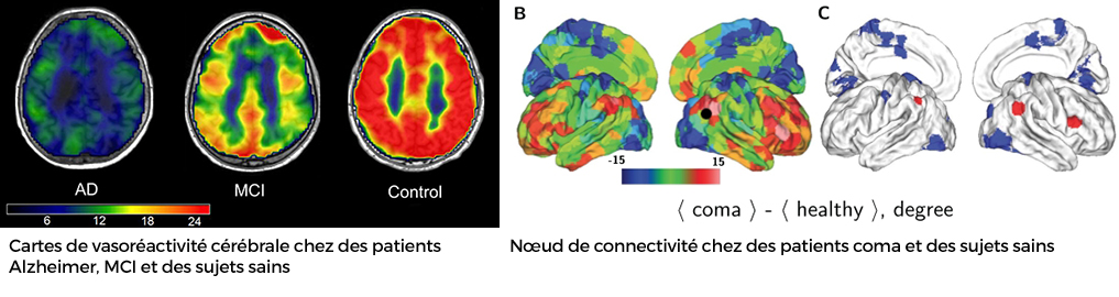
Achard S, Delon-Martin C, Vértes PE, Renard F, Schenck M, Schneider F, Heinrich C, Kremer S, Bullmore ET. Hubs of brain functional networks are radically reorganized in comatose patients. Proc Natl Acad Sci U S A (PNAS). 2012. 109(50):20608-13
Cantin S, Villien M, Moreaud O, Tropres I, Keignart S, Chipon E, Le Bas JF, Warnking J, Krainik A. Impaired cerebral vasoreactivity to CO(2) in Alzheimer's disease using BOLD fMRI. Neuroimage 2011;58(2):579-587.
Membres de l'équipe
- Sophie ACHARD
- Cleopha AOV
- Emmanuel BARBIER
- Antoine BARRIER
- Camille BEETS
- Pierre BOUZAT
- Anna CASTRIOTO
- Thomas CHRISTEN
- Nora COLLOMB
- Thomas COUDERT
- Clothilde COURIVAUD
- Bettina DEBU
- Chantal DELON-MARTIN
- Antoine DEPAULIS
- Olivier DETANTE
- Michel DOJAT
- Jean-Come DOUTEAU
- Bayan EL AMINE
- Florence FAUVELLE
- Valérie FRAIX
- Paul GALLOUX
- Tobias GAUSS
- Sylvie GRAND
- Marie GUILMAIN
- Chiara HARTMANN
- Adrian KASTLER
- Alexandre KRAINIK
- Loïc LEGRIS
- Marion LE MARECHAL
- Benjamin LEMASSON
- Hervé MATHIEU
- Sara MEONI
- Razan MHANNA
- Vanille MILLASSEAU
- David MINLOUBOU
- Olivier MONTIGON
- Elena MORO
- Geoffroy OUDOUMANESSAH
- Jean-Francois PAYEN DE LA GARANDERIE
- Pierre PELISSIER
- Elodie PIOT
- Sébastien RIGOLLET
- Claire ROME
- Marc SAGHIAH
- Romane SALINGUE
- Loan SAMALENS
- Emmanuelle SCHMITT
- Maité SILVA MARTINS MARCAL
- Vasile STUPAR
- Vaëa TESAN
- Jan WARNKING