
Directors : Benjamin LEMASSON and Thomas CHRISTEN
Themes of research
The research field of the team is in vivo biomedical magnetic resonance imaging (MRI). Our aims are the development, the validation and the application of in vivo MRI methods in the field of the clinical, biological and cognitive neuroscience. Since 2012, the team and the MRI facility IRMaGe are members of the national infrastructures 'France Life Imaging (FLI)'. The studies are performed on a preclinical and a clinical levels. They imply the coexistence of two overlapping components, one centered on the development of methods, the other on their evaluation. This duality is reflected in the multidisciplinary backgrounds of the members of the team, from physics to life sciences. More specifically, we address:
-
Development of functional and physiological imaging
We develop new imaging tools to characterize brain microvascularization: vessel size index (VSI), permeability of the vessel wall, blood volume (CBV) and blood flow (CBF), tissue oxygen saturation (StO2, quantitative BOLD), and cerebrovascular vasoreactivity. Based on these physiological information, we develop advanced functional MRI (fMRI) technique (e.g. joint detection estimation, in collaboration with Inria and Neurospin) and functional connectivity MRI (e.g. graph-based approach, in collaboration with GIPSA). We pursue these developments to improve the use of fMRI in patients. Our expertise in fMRI serves also numerous collaborations in cognitive neuroscience (visual, motor, auditory and olfactory systems) and neurology (brain tumor, stroke, coma, brain trauma, aging). -
Development of imaging biomarker
To exploit the structural, metabolic and perfusion data collected with MRI, we develop innovative tools for automated information extraction. We developed the approach LOCUS, based on Markovien models in a Baysian framework, to automatically segment tissue structures and brain lesions. We also exploit multiparametric MRI to obtain fingerprints of healthy and diseased tissues (coll. Inria). -
Imaging-guided therapy
Based on MRI, we evaluate advanced therapeutic strategies, such as microbeam X-ray therapy or anti-angiogenic therapies against brain tumors, stem cells therapy in the context of stroke and anti-oedematous therapy in the context of brain trauma. Since 2012, the team is par of the national infrastructure ECELL FRANCE, which supports the development of the therapeutic strategies based on adult mesenchymal stem cells.
Some tools developed by the team are available:
-
DCESIM
- P-LOCUS (in French)
Partners 
Thesis of the team (in french)
Publications
S. Hamelin, V. Stupar, L. Mazière, J. Guo, W. Labriji, C. Liu, L. Bretagnolle, S. Parrot, E. L. Barbier, A. Depaulis, F. Fauvelle. In vivo GABA increase as a biomarker of the epileptogenic zone: an unbiased metabolomics approach. Epilepsia, 62(1):163-175, 2021.
P. Roca, A. Attyé, L. Colas, A. Tucholka, P. Rubini, S. Cackowski, J. Ding, J.-F. Budzik, F. Renard, S. Doyle, E. L. Barbier, I. Bousaid, R. Casey, S. Vukusic, N. Lassau, S. Verclytte, F. Cotton; OFSEP Investigators;Steering Committee; Investigators; Imaging group. Artificial intelligence to predict clinical disability in patients with multiple sclerosis using FLAIR MRI. Diagnostic and Interventional Imaging. 101(12):795-802, 2020.
T. Deruelle, F. Kober, A. Perles-Barbacaru, T. Delzescaux, V. Noblet, E. L Barbier, M. Dojat. A Multicenter Preclinical MRI Study: Definition of Rat Brain Relaxometry Reference Maps. Frontiers in Neuroinformatics, 14:22, 2020.
L. Hirschler, N. Collomb, J. Voiron, S. Köhler, E. L. Barbier, J. M. Warnking. SAR comparison between CASL and pCASL at high magnetic field and evaluation of the benefit of a dedicated labeling coil. Magnetic Resonance in Medicine 83(1):54-261, 2020
G. J.-P. C. Becq, T. Habet, N. Collomb, M. Faucher, C. Delon-Martin, V. Coizet, S. Achard, E. L. Barbier. Functional connectivity is preserved but reorganized across several anesthetic regimes. Neuroimage, 219:116945, 2020.
G. J.-P. C. Becq, E. L. Barbier, S. Achard. Brain networks of rats under anesthesia using resting-state fMRI: comparison with dead rats, random noise and generative models of networks. Journal of Neural Engineering, 17(4):045012, 2020.
C. Verry, S. Dufort, B. Lemasson, S. Grand, J. Pietras, I. Troprès, Y. Crémillieux, F. Lux, S. Mériaux, B. Larrat, J. Balosso, G. Le Duc, E. L. Barbier, O. Tillement. Targeting brain metastases with ultrasmall theranostic nanoparticles, a first-in-human trial from an MRI perspective. Science Advances, 6(29), eaay5279, 2020.
F. Natali, C. Dolce, J. Peters, C. Stelletta, B. Demé, J. Ollivier, M. Boehm, G. Leduc, I. Piazza, A. Cupane, E. L. Barbier. Anomalous water dynamics in brain: a combined diffusion Magnetic Resonance Imaging and Neutron Scattering investigation. Journal of the Royal Society Interface 16(157):20190186, 2019.
Hirschler L, Debacker CS, Voiron J, Köhler S, Warnking JM, Barbier EL. Interpulse phase corrections for unbalanced pseudo-continuous arterial spin labeling at high magnetic field. Magn Reson Med. 2018 Mar;79(3):1314-1324. doi: 10.1002/mrm.26767.
Dojat M, Pizzagalli F, Hupé JM. Magnetic resonance imaging does not reveal structural alterations in the brain of grapheme-color synesthetes. PLoS One. 2018 Apr 4;13(4):e0194422. doi: 10.1371/journal.pone.0194422.
Kragel PA, Kano M, Van Oudenhove L, Ly HG, Dupont P, Rubio A, Delon-Martin C, Bonaz BL, Manuck SB, Gianaros PJ, Ceko M, Reynolds Losin EA, Woo CW, Nichols TE, Wager TD. Generalizable representations of pain, cognitive control, and negative emotion in medial frontal cortex. Nat Neurosci. 2018 Feb;21(2):283-289. doi: 10.1038/s41593-017-0051-7.
R. He, A. Moisan, O. Detante, C. Rémy, A. Krainik, E. L. Barbier, B. Lemasson. Evaluation of parametric response mapping to assess therapeutic response to human mesenchymal stem cells after experimental stroke. Cell Transplantation 26(8) 1462-1471, 2017.
K. Pernet Gallay, P. H. Jouneau, J. Delaroche, R. Farion, C. Rémy, E. L. Barbier. Vascular permeability in the RG2 glioma model can be mediated by macropinocytosis and be independent of the opening of the tight junction. Journal of Cerebral Blood Flow and Metabolism, 37(4):1264–1275, 2017.
A. Moisan, I Favre, C. Rome, F. de Fraipont, E. Grillon, N. Coquery, H. Mathieu, V. Mayan, B. Naegelé, M. Hommel, M.-J. Richard, E. L. Barbier, C. Rémy, O Detante. Intravenous injection of clinical grade human MSC after experimental stroke: functional benefit and microvascular effect. Cell transplantation, 12:2157-2171, 2016.
B. Lemasson, N. Pannetier, N. Coquery, Ligia S. B. Boisserand, Nora Collomb, N. Schuff, M. Moseley, G. Zaharchuk, E.L. Barbier, T. Christen. MR Vascular Fingerprinting in Stroke and Brain Tumors Models. Scientific Reports, 6:37071, 2016.
Bellot E, Coizet V, Warnking J, Knoblauch K, Moro E, Dojat M. Effects of aging on low luminance contrast processing in humans. Neuroimage. 2016 Oct 1;139:415-426. doi: 10.1016/j.neuroimage.2016.06.051.
A. Millet, P. Bouzat, C. Batandier, A. Daoust, R. Farion, L. Gaide-Chevronnay, E. L. Barbier, T. Debillon, E. Fontaine, J.-F. Payen. Erythropoietin and its derivates modulate mitochondrial dysfunction after diffuse traumatic brain injury. J. Neurotrauma 33(17):1625-1633, 2016.
N. Coquery, V. Stupar, R. Farion, S. Maunoir-Regimnal, E. L. Barbier, C. Rémy, F. Fauvelle. The three glioma rat models C6, F98 and RG2 exhibit different metabolic profiles: in vivo 1H MRS and ex vivo 1H HRMAS combined with multivariate statistics. Metabolomics, 11:1834–1847, 2015.
Ramanoël S, Kauffmann L, Cousin E, Dojat M, Peyrin C. Age-Related Differences in Spatial Frequency Processing during Scene Categorization. PLoS One. 2015 Aug 19;10(8):e0134554. doi: 10.1371/journal.pone.0134554. eCollection 2015.
B. Lemasson, A. Bouchet, C. Maisin, T. Christen, G. Le Duc Géraldine, C. Remy, E. L. Barbier, R Serduc. Multiparametric MRI as an early biomarker of individual therapy effect during concomitant treatment of brain tumors. NMR in Biomed, 28(9):1163-73, 2015.
Menze BH, Jakab A, Bauer S, Kalpathy-Cramer J, Farahani K, Kirby J, Burren Y, Porz N, Slotboom J, Wiest R, Lanczi L, Gerstner E, Weber MA, Arbel T, Avants BB, Ayache N, Buendia P, Collins DL, Cordier N, Corso JJ, Criminisi A, Das T, Delingette H, Demiralp Ç, Durst CR, Dojat M, Doyle S, Festa J, Forbes F, Geremia E, Glocker B, Golland P, Guo X, Hamamci A, Iftekharuddin KM, Jena R, John NM, Konukoglu E, Lashkari D, Mariz JA, Meier R, Pereira S, Precup D, Price SJ, Raviv
TR, Reza SM, Ryan M, Sarikaya D, Schwartz L, Shin HC, Shotton J, Silva CA, Sousa N, Subbanna NK, Szekely G, Taylor TJ, Thomas OM, Tustison NJ, Unal G, Vasseur F, Wintermark M, Ye DH, Zhao L, Zhao B, Zikic D, Prastawa M, Reyes M, Van Leemput K. The Multimodal Brain Tumor Image Segmentation Benchmark (BRATS). IEEE Trans Med Imaging. 2015 Oct;34(10):1993-2024. doi: 10.1109/TMI.2014.2377694.
J. Bouvier, O Detante, F. Tahon, A. Attye, T. Perret, D. Chechin, M. Barbieux, K. Boubagra, K. Garambois, I. Tropres, S. Grand, E. L. Barbier, A. Krainik. Reduced CMRO2 and cerebrovascular reserve in patients with severe intracranial arterial stenosis: a combined multiparametric qBOLD oxygenation and BOLD fMRI study. Human Brain Mapping, 36(2):695-706, 2015.
A. Daoust, S. Bohic, Y. Saoudi, C. Debacker, S. Gory-Fauré, A. Andrieux, E. L. Barbier, J.-C. Deloulme. Neuronal transport defects of the MAP6 KO mouse – a model of schizophrenia – and alleviation by Epothilone D treatment, as observed using MEMRI. Neuroimage. 96C:133-142, 2014
T. Christen, P. Bouzat, N. Pannetier, N. Coquery, A. Moisan, B. Lemasson, S. Thomas, E. Grillon, O. Detante, C. Rémy, J.-F. Payen, E.L. Barbier. Tissue oxygen saturation mapping with magnetic resonance imaging. Journal of Cerebral Blood Flow and Metabolism. 34(9):1550-7, 2014
Hupé J, Bordier C, Dojat M. A BOLD signature of eyeblinks in the visual cortex Neuroimage 2012;61(1):149-161
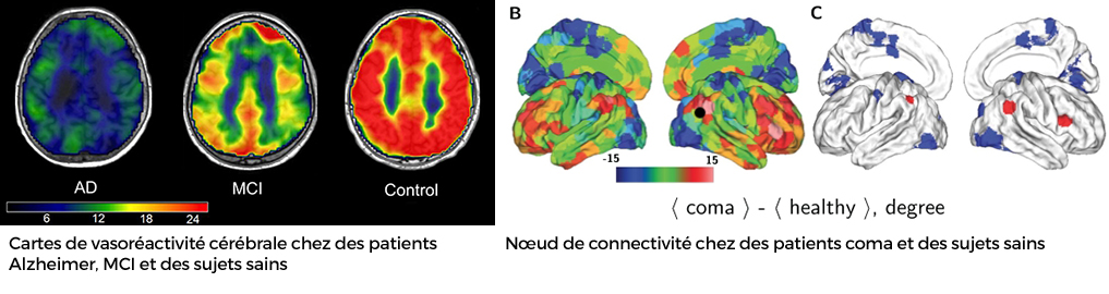
Achard S, Delon-Martin C, Vértes PE, Renard F, Schenck M, Schneider F, Heinrich C, Kremer S, Bullmore ET. Hubs of brain functional networks are radically reorganized in comatose patients. Proc Natl Acad Sci U S A (PNAS). 2012. 109(50):20608-13
Cantin S, Villien M, Moreaud O, Tropres I, Keignart S, Chipon E, Le Bas JF, Warnking J, Krainik A. Impaired cerebral vasoreactivity to CO(2) in Alzheimer's disease using BOLD fMRI. Neuroimage 2011;58(2):579-587.
Members
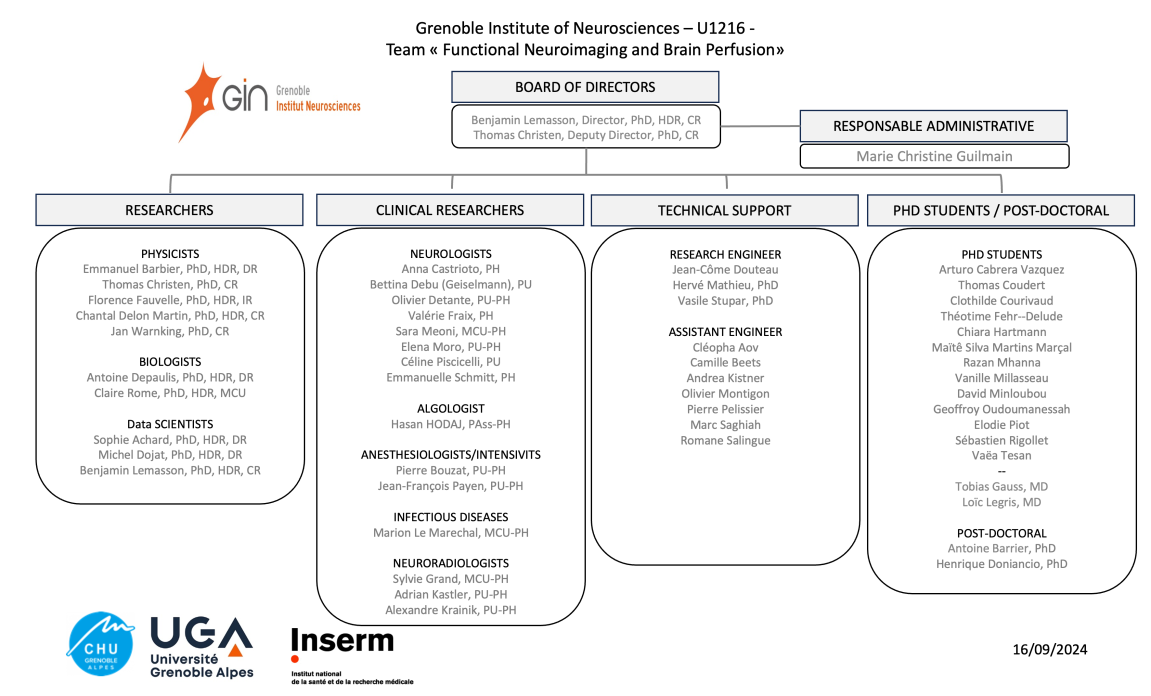
- Sophie ACHARD
- Cleopha AOV
- Emmanuel BARBIER
- Antoine BARRIER
- Camille BEETS
- Pierre BOUZAT
- Anna CASTRIOTO
- Thomas CHRISTEN
- Nora COLLOMB
- Thomas COUDERT
- Clothilde COURIVAUD
- Bettina DEBU
- Chantal DELON-MARTIN
- Antoine DEPAULIS
- Olivier DETANTE
- Michel DOJAT
- Jean-Come DOUTEAU
- Bayan EL AMINE
- Florence FAUVELLE
- Valérie FRAIX
- Paul GALLOUX
- Tobias GAUSS
- Sylvie GRAND
- Marie GUILMAIN
- Chiara HARTMANN
- Adrian KASTLER
- Alexandre KRAINIK
- Loïc LEGRIS
- Marion LE MARECHAL
- Benjamin LEMASSON
- Hervé MATHIEU
- Sara MEONI
- Razan MHANNA
- Vanille MILLASSEAU
- David MINLOUBOU
- Olivier MONTIGON
- Elena MORO
- Geoffroy OUDOUMANESSAH
- Jean-Francois PAYEN DE LA GARANDERIE
- Pierre PELISSIER
- Elodie PIOT
- Sébastien RIGOLLET
- Claire ROME
- Marc SAGHIAH
- Romane SALINGUE
- Loan SAMALENS
- Emmanuelle SCHMITT
- Maité SILVA MARTINS MARCAL
- Vasile STUPAR
- Vaëa TESAN
- Jan WARNKING