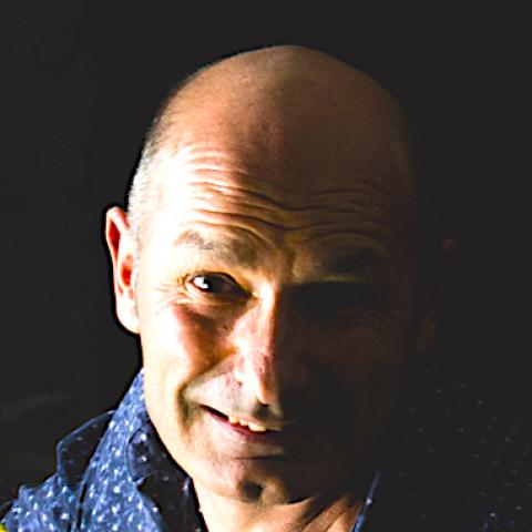
MICHEL DOJAT
Directeur de recherche (INSERM)
- Share
- Share on Facebook
- Share on X
- Share on LinkedIn
Eq B.Lemasson/T.Christen

Contact details
Building : Grenoble Institut des Neurosciences
Office : GIN R/074
Michel.Dojat@univ-grenoble-alpes.fr
Social networks :
Research topics
Neuroinformatics, Neuroimaging, Signal and Image Analysis, Human Vision
I am involved in human and animal brain studies based on functional neuroimaging. My favorite application is Human Vision (color vision, color assimilation grapheme-color synesthesia and saccadic eye movements). I also study vision deficits especially related to Parkinson's disease progression and Blepharospasm.
At the methodological level, I develop methods for MR brain image analysis: brain lesion segmentation (e.g. RADIOAIDE sponsored by ANR (2022-26), brain connectivity), functional brain connectivity (GRAACE project, (2020-23) sponsored by the MIAI Grenoble Institute), and biomarker imaging extraction (DAISIES project, 2022-2025) sponsored by the AURA region and MIAI Grenoble Institute.
I manage projects in Neuroinformatics to facilitate data and image processing sharing in neuroimaging (see the precursors NeuroLOG and Shanoir) and the current IAM (Image Analysis and Management) project for designing a versatile infrastructure for data and processing tool management for in vivo imaging in the context of France Life Imaging. See the recent RT MSSEG on MS lesion detection, the RT APPNING or the SAIN research network about animal population imaging. Interoperability between FLI-IAM and others life science platforms is studied via MUDI4LS project and EBRAINS.
I am Deputy scientific director for digital biology and digital medicine at Inria and member of Statify. I am one of the cofounders of PIXYL, a start-up that develops innovative computerized solutions for medical research. I am member of the direction committee of IXXI, a research structure on complex systems. I participate to the thinking group CARMEN (Conscience Attention and Représentation Mentale).
I also worked on AI-based systems for patient monitoring, temporal reasoning and models for medical reasoning. The application field was automatic control of therapy delivered to patients hospitalized in intensive care units. A prototype of a computerized protocol for pressure support delivrance, NéoGanesh was firstly designed. Several scientific publications demonstrate its therapeutic interest and an industrial version embedded into a commercial respirator in now available SmartCARE/PS (see details).
Teaching
Biophysique et imagerie médicale, INFORMATIQUE SCIENTIFIQUE, Neurosciences, Radiologie et imagerie médicale, TRAITEMENT INFORMATION
Lectures
- 2025
Digital Health in the 21th (The 4th Annecy Round Table on CPR, Jean-Christophe Richard, Apr 2025, Annecy, France) - 2024
The multi-dimensional aspect of uncertainty - 2023
Superior Colliculus in Parkinson Disease - 2021
FLI-IAM* - 2018
AI&Neurosciences
Vision-MR2:
- Color Vision BookChapter
- Human Vision Lecture 2025
TIS-2017: Medical Image Processing Introduction, MRI, Image Processing
TIS-2025: Imaging & Machine Learning PartI-1, PartI-2, PartII-1, PartII-2
MRI for neuroscientists: Intro, Stats, Image Processing, Segmentation, MVPA
Curriculum vitae
I was born in France in 1959. I received my engineering diploma from Institut National des Sciences Appliquées de Lyon in applied physics in 1982, my PhD thesis in Computer Science in 1994 from University de Compiègne and my 'Habilitation à Diriger des Recherches' in "Medical Informatics" in 1999 from University J. Fourier de Grenoble. From 1985 to 1988, I worked as research engineer in Télémécanique Electrique (now Schneider Electric) research center (Nanterre, 92, Fr) on the modeling and design of sensors for industrial process control. Since 1988, I am with INSERM. During 10 years (1988-1998), I worked in the INSERM U296 at Henri Mondor Hospital (Créteil, Fr), a Laboratory devoted to Respiratory Physiology, where I developed AI-oriented systems for supervision artificial ventilation. During this period, I was member of the AI lab (LAFORIA) of University Paris 6. In 1998, I joined the functional neuroimaging team (formely U594) at the Grenoble Institut des Neurosciences (U1216 ex-U836). I co-founded Pixyl, a Inserm-Inria start-up devoted to AI for medical imaging. I am Deputy scientific director for digital biology and digital medicine at Inria since 2023.

Publications
List of available publications :
via Orcid.
via Publons.
via GoogleScolar.
via Loops.
via Hal.
En imagerie médicale, faire de l'IA une alliée (Le Monde "Science & Médecine" 01/06/2022)
L'intelligence artificielle en médecine - promesses, limites et enjeux (Futurologie, 15/04/2025)
Bibliographic search
Other links
- Share
- Share on Facebook
- Share on X
- Share on LinkedIn