
- Share
- Share on Facebook
- Share on LinkedIn
Communiqué / Team I.Arnal/A.Andrieux
On July 23, 2025
By studying the MAP6d1 protein, a team of researchers at GIN has made important discoveries in understanding the molecular mechanisms governing microtubule doublet formation and cilia physiology.
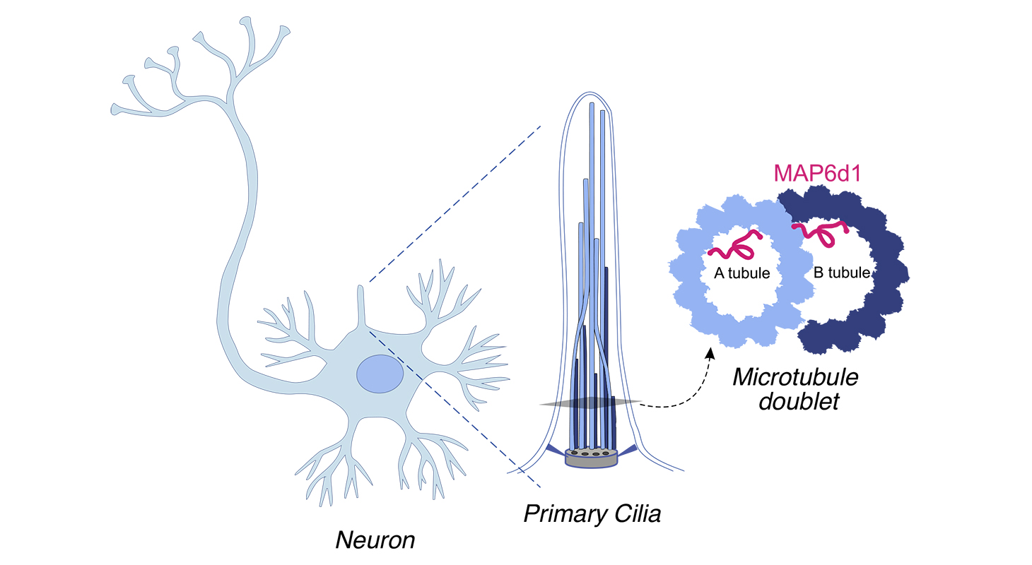
Most eukaryotic cells possess cilia that serve vital functions. Flagella and motile cilia are responsible for moving cells, while primary cilia, known as non-motile, function as small antennae involved in sensory perception and signal transduction. Defects in cilia assembly and structure can result in a number of diseases, collectively known as ciliopathies.
Microtubules in the axonemes of cilia and flagella form complex arrays of microtubule doublets, ensuring the integrity and function of these cellular appendages. However, the mechanisms by which microtubule doublets are assembled remain largely unknown.
Researchers from the “Dynamics and Structure of the Neuronal Cytoskeleton” team led by Isabelle Arnal and Annie Andrieux have focused their attention on MAP6d1, a brain-specific protein that contains motifs targeting the lumen of microtubules, i.e., the interior space of the hollow cylinder formed by the assembly of tubulin protofilaments.
By combining in vitro reconstitution, real-time fluorescence microscopy techniques, and cryo-electron tomography, they uncovered a direct role for MAP6d1 in the assembly and stabilization of microtubule doublets. MAP6d1 stands out for it ability to spontaneously assemble microtubule doublets, a process dependent on its microtubule-luminal binding sites.
Remarkably, MAP6d1 also induces the formation of protofilaments within the microtubule lumen, a previously undescribed phenomenon suspected to enhance the mechanical resistance of microtubules.
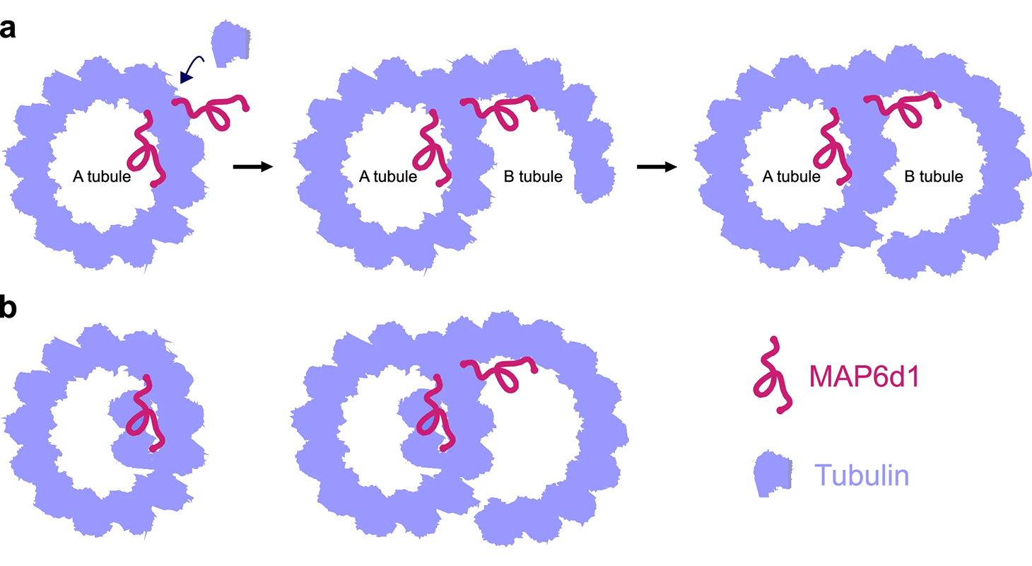
a MAP6d1 binds to the A-tubule lattice, recruits free tubulin dimers to initiate the B-tubule and forms a doublet microtubule. b During co-polymerisation with microtubules, MAP6d1 assembles protofilaments in the lumen.
Another significant finding is that MAP6d1 localizes to the primary cilia of cultured mouse neurons, where it is involved in maintaining their architecture. Its postnatal expression suggests a key role in the elongation and progressive maturation of cilia, a process that lasts for several weeks after birth.
Together, these results represent a major advance in our understanding of the molecular mechanisms governing the formation of microtubule doublets and ciliary physiology.
Reference :
The Mn-motif protein MAP6d1 assembles ciliary doublet microtubules.
Dharshini Gopal, Juliette Wu, Julie Delaroche, Christophe Bosc, Manon De Andrade, Eric Denarier, Gregory Effantin, Annie Andrieux, Sylvie Gory-Fauré, Laurence Serre & Isabelle Arnal
Nature Communications, 2025, 16 (1), pp.6210. (10.1038/s41467-025-61679-0)
Date
- Share
- Share on Facebook
- Share on LinkedIn