
- Share
- Share on Facebook
- Share on LinkedIn
Communiqué / Alzheimer, Team A.Buisson, Research
On October 8, 2026
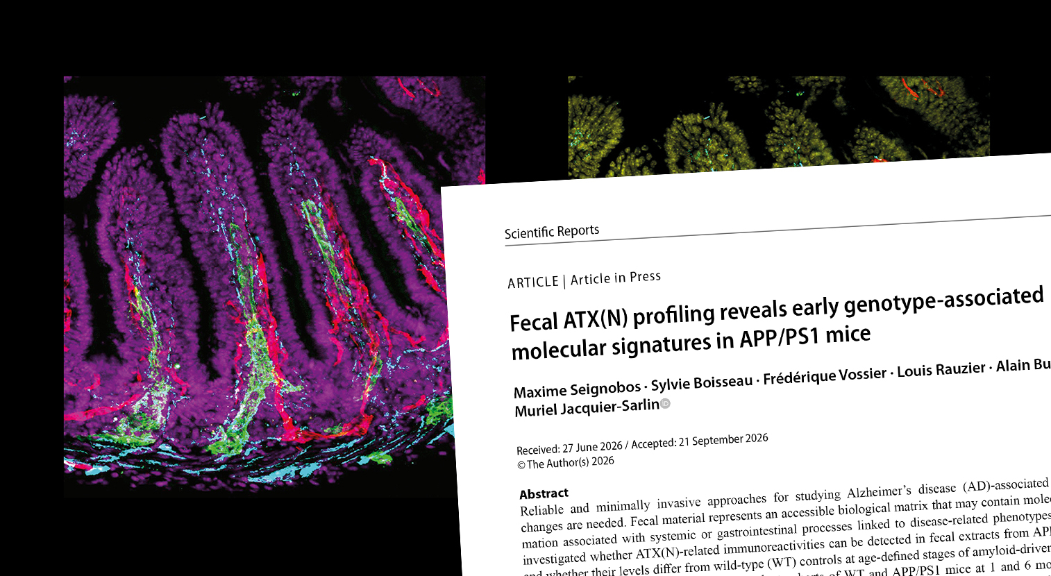
A study conducted at the Grenoble Institute of Neurosciences shows that several proteins associated with Alzheimer's disease can be detected in mouse stool samples and that some of these markers show changes at a very early stage, even before the amyloid plaques characteristic of the disease become detectable in the brain. Published in Scientific Reports, these findings open up a new avenue of research towards simple, non-invasive testing methods, whose relevance must now be evaluated in humans.
Searching for more accessible biomarkers
Today, brain imaging and cerebrospinal fluid analysis following a lumbar puncture are among the reference examinations used to diagnose Alzheimer's disease. However, these procedures remain costly and burdensome.
“We became interested in stool samples as a complementary biological matrix for studying Alzheimer's disease. Easy to access, inexpensive and simple to collect, they could provide additional information alongside that obtained from cerebrospinal fluid or blood when analysing certain biomarkers,” explains Maxime Seignobos, who conducted his doctoral research at the GIN within the “Neuropathologies and Synaptic Dysfunctions” team, under the supervision of Muriel Jacquier-Sarlin.
His research focused on several proteins associated with Alzheimer's disease: amyloid-beta, different forms of the Tau protein, as well as markers of neurodegeneration, synaptic function and inflammation.
The researchers compared the signals detected in stool samples from APP/PS1 mice, a model of Alzheimer's disease, with those observed in control mice at one and six months of age.
Differences detectable at a very early stage
The first finding was that amyloid-beta and Tau protein could be detected in stool samples, including those from control mice. Their presence alone is therefore not a sign of disease.
However, differences were observed between the two groups.
As early as one month of age, several forms of amyloid-beta and phosphorylated Tau showed higher signals in APP/PS1 mice. The same was true for NfL, a protein used as a marker of neurodegeneration, and synaptophysin, a protein associated with synaptic function.
“What is interesting is that we see this increase in the stool samples of APP/PS1 mice at a fairly early stage, as early as one month of age, before amyloid deposits appear in the brain,” points out Maxime Seignobos.
Indeed, in this mouse model of Alzheimer's disease, the first amyloid plaques only become detectable in the cortex from three months of age. The changes observed in stool samples therefore precede them by several weeks.
What is the link between the brain and the gut?
How do these biomarkers end up in mouse stool samples? This initial study cannot yet provide an answer. The detected proteins could have different origins, and their presence in stool does not necessarily mean that they come directly from the brain.
Understanding the origin of these signals and the mechanisms that might link the brain and the gut is precisely the focus of the second part of Maxime Seignobos's doctoral research, which is expected to be the subject of a forthcoming publication.
These initial findings in mice represent “the first step that paves the way for the clinical study”, the young researcher explains.
A clinical study underway
The transition to human research is already underway through the FECALZ project. A clinical study conducted in collaboration with Grenoble Alpes University Hospital and the Grenoble Arc Alpin Memory Resources and Research Centre (CMRR) has enrolled 135 volunteers: healthy individuals, people with mild cognitive impairment and patients with Alzheimer's disease. Analysis of their stool samples will help determine whether the differences observed in mice are also found in humans.
“In the long term, the aim would be to contribute to the development of diagnostic tools enabling earlier and more accessible detection of the disease,” concludes Maxime Seignobos.
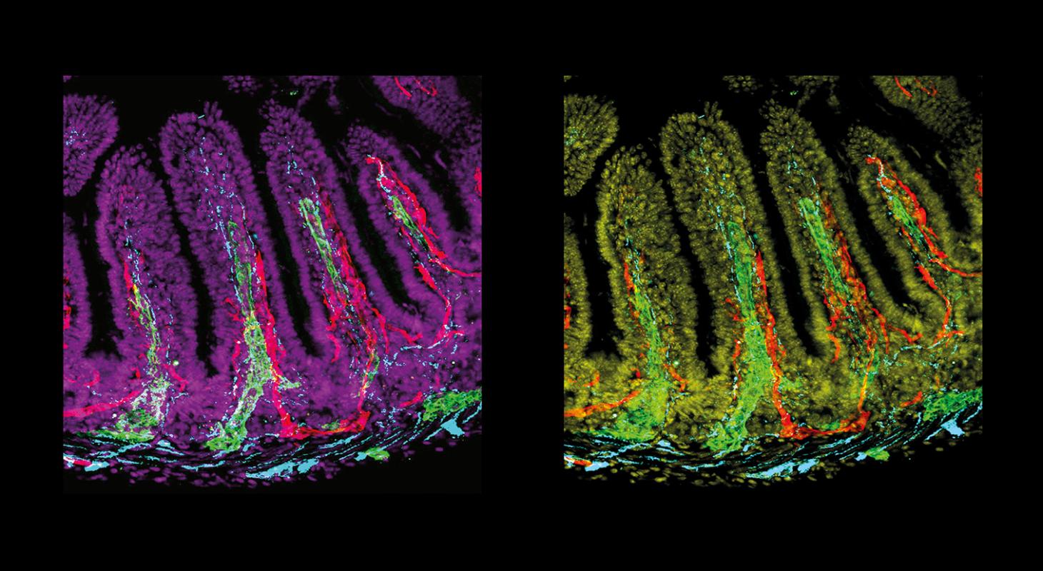
Fluorescence microscopy images showing intestinal villi, structures involved in nutrient absorption. Different fluorescent markers allow the visualization of Tau protein (cyan), vascular networks, and other structures within the intestinal tissue. The origin and fate of proteins associated with Alzheimer's disease in the intestine are being investigated in further research conducted by Maxime Seignobos as part of his PhD.
Reference :
Fecal ATX(N) profiling reveals early genotype-associated molecular signatures in APP/PS1 mice.
Seignobos M., Boisseau S., Vossier F., Rauzier L., Buisson A. & Jacquier-Sarlin M.
Scientific Reports, 2026.
Date
- Share
- Share on Facebook
- Share on LinkedIn