
- Share
- Share on Facebook
- Share on LinkedIn
Communiqué / Team A.Buisson
From July 16, 2026 to September 30, 2026
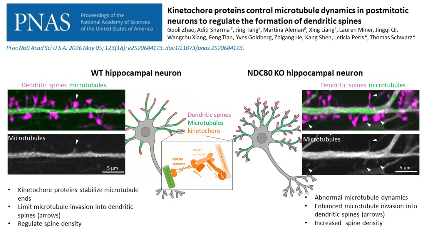
Essential for cell division, kinetochore proteins also play an unexpected role in mature neurons. While a US research team had observed their influence on dendritic spine formation, Leticia Peris’s expertise helped uncover the underlying mechanism: these proteins control microtubule dynamics within neurons.
Kinetochore proteins in neurons
The kinetochore is best known for its role in cell division. This protein complex enables microtubules to attach to chromosomes, ensuring that they are correctly distributed between the two daughter cells. At first glance, this function seems far removed from the physiology of neurons, which no longer divide once they have differentiated.
However, several kinetochore proteins remain present in neuronal processes after cell division has ended. What role might they play? This was the question investigated by Thomas Schwarz, a researcher at Boston Children’s Hospital and Harvard Medical School.
In neurons lacking certain kinetochore proteins, including NDC80 or DSN1, his team observed a marked increase in dendritic spine density, both in cultured mouse neurons grown and in the mouse cortex. These tiny protrusions host the majority of excitatory synapses and therefore play an essential role in communication between neurons.
To understand this phenomenon, the US team wanted to observe the behaviour of microtubules within dendritic spines in real time.
Following a recommendation from Christophe Leterrier, a researcher at the Institute of Neurophysiopathology in Marseille, Thomas Schwarz contacted Leticia Peris, a researcher at the Grenoble Institute of Neuroscience whose expertise in microtubule dynamics is internationally recognised.
“Our hypothesis was that the increase in spine density might be linked to increased microtubule invasion of these structures,” explains Leticia Peris.
Observing microtubules in real time
To test this hypothesis, the US team sent cryopreserved mouse neurons from the United States to Grenoble, together with the genetic tools needed to remove or reintroduce the NDC80 protein. This innovative preservation method enabled Leticia Peris to work directly with the cells engineered in Boston.
Together with Aditi Sharma and Martina Aleman, Leticia Peris designed and conducted a series of experiments to observe dendritic spines and the growing ends of microtubules simultaneously. Using two fluorescent markers and spinning-disk confocal microscopy, the scientists were able to track microtubules as they left the dendrite and transiently entered a spine.
The results confirmed the researcher’s hypothesis. In the absence of NDC80, microtubules became more dynamic and invaded dendritic spines more frequently. Over a five-minute observation period, the proportion of spines visited by a microtubule was more than twice that measured in control neurons. This increase was accompanied by a 38% rise in spine density.
But the experiment went one step further. When the scientists reintroduced normal NDC80 into the neurons, both the frequency of microtubule entry into the spines and spine density returned to their original levels. By contrast, a version of NDC80 lacking the region that allows it to bind to microtubules did not produce this effect.
“This result really changed the scope of the study,” Leticia Peris emphasises. “It shows that NDC80 does not merely play an indirect role in dendritic spine formation: its ability to interact with microtubules is the determining factor.”
A brake on dendritic spine formation
In mature neurons, kinetochore proteins therefore appear to act as a brake. By stabilising microtubule ends, they limit their entry into dendritic spines and, consequently, the formation or maintenance of these structures. When these proteins are absent, microtubules become more dynamic, enter the spines more frequently and spine density increases.
This discovery reveals a remarkable continuity between two functions that initially seemed entirely unrelated. During cell division, as in mature neurons, kinetochore proteins interact with microtubule ends. However, this same property serves two very different purposes: distributing chromosomes in one case and helping to organise neuronal connections in the other.
This reuse of the same molecular machinery in two very different contexts illustrates how living organisms can repurpose existing tools for new functions.
Reference:
Kinetochore proteins control microtubule dynamics in postmitotic neurons to regulate the formation of dendritic spines
Zhao G, Sharma A, Tang J, Aleman M, Liang X, Miner L, Qi J, Xiang W, Tian F, Goldberg Y, He Z, Shen K, Peris L, L Schwarz T.
Proc Natl Acad Sci U S A. 2026 May 5;123(18):e2520684123. doi: 10.1073/pnas.2520684123. Epub 2026 Apr 27. PMID: 42044343; PMCID: PMC13132258.
Date
- Share
- Share on Facebook
- Share on LinkedIn