
- Share
- Share on Facebook
- Share on LinkedIn
Communiqué / Team A.Buisson
On November 2, 2026
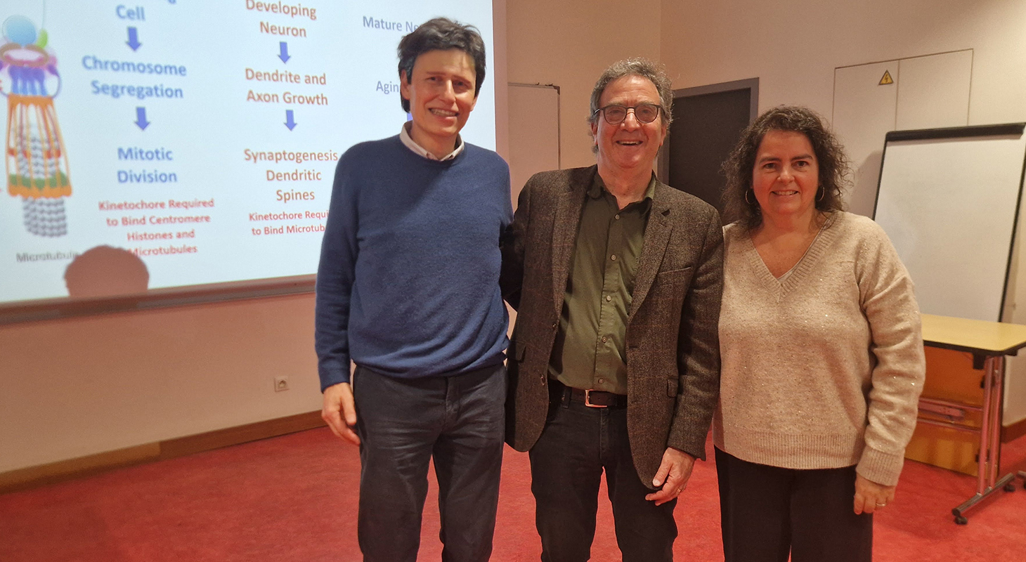
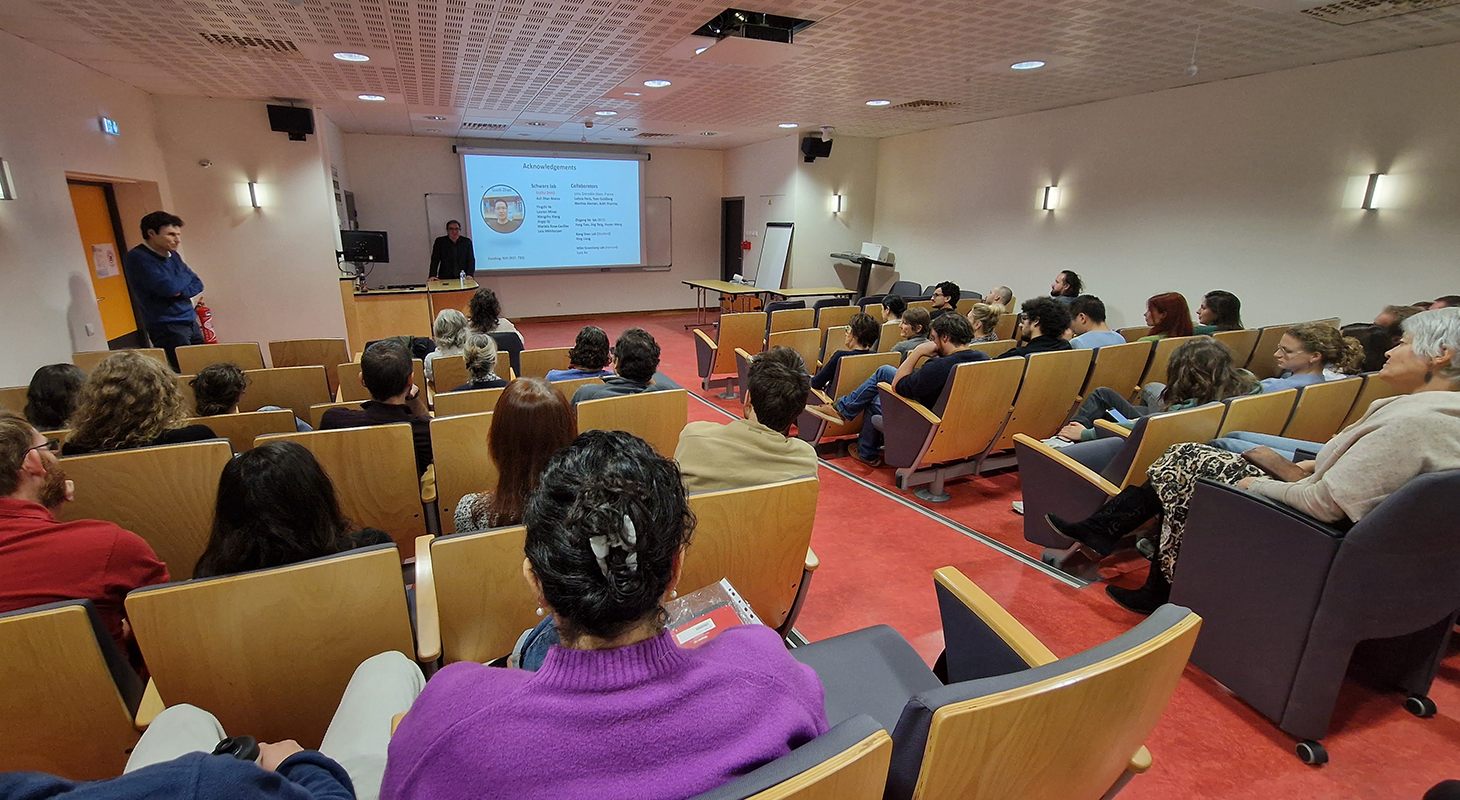
How can a structure previously known for its role in cell division be involved in synapse formation and neuronal survival? During a seminar at the Grenoble Institut des Neurosciences on October 30, 2026, Thomas Schwarz, a researcher at Harvard, recounted the scientific investigation that led him to uncover these unexpected functions, with a crucial contribution from Leticia Peris.
An Unexpected Discovery
The kinetochore is a protein complex located at the centromere of chromosomes. It connects chromosomes to microtubules and enables their separation during cell division. “The kinetochore had no known function outside mitosis and meiosis,” Thomas Schwarz recalled at the beginning of his talk. “Nobody had studied it in the nervous system. Why would they? Neurons, as everyone knows, do not divide.”
Yet, while investigating the mechanisms governing synapse formation and maturation in fruit flies (Drosophila), Thomas Schwarz and his team made a surprising discovery. Searching for genes involved in this process, they identified a mutation that disrupted the development of neuromuscular junctions. The culprit? MIS12, a gene encoding a kinetochore protein!
“I thought, Guoli had made a mistake in his genetic experiments. This can't be right!” Schwarz recalled, describing his reaction when his postdoctoral researcher Guoli Zhao first reported the results.
But there was no mistake…
To confirm their discovery, the team turned its attention to other kinetochore proteins. Almost all the mutations they tested produced similar abnormalities in neuromuscular junction development.
The evidence was mounting.
From Fruit Flies to Mammalian Neurons
Increasingly intrigued, the researchers extended their investigation to mammals. By reducing the expression of several kinetochore proteins, including MIS12, DSN1 and NDC80, in rodent neurons, they observed striking changes in neuronal morphology: axons grew longer, growth cones became larger and, most notably, dendritic spines increased in number. These small protrusions on dendrites are the sites of most excitatory synapses.
But how could proteins known for their role in cell division influence neuronal morphology?
Another player emerged: microtubules, components of the cytoskeleton that help organize neurons and support their growth. Indeed, the researchers found that reducing the levels of certain kinetochore proteins increased microtubule dynamics.
“At that point, we were wondering what any of this had to do with dendritic spine formation,” Schwarz explained.
A Crucial Collaboration with Grenoble
The investigation then took a new turn following an encounter. At a conference, a colleague suggested that Thomas Schwarz contact Leticia Peris, a researcher at the Grenoble Institut des Neurosciences who specializes in microtubules within the synaptic compartment.
For many years, Leticia Peris has studied microtubule dynamics and their role in the stability of neuronal connections. In particular, she investigates their transient entry into dendritic spines.
The collaboration began. Drawing on her expertise and the imaging techniques available at the GIN, Leticia Peris, together with her PhD students Aditi Sharma and Martina Aleman and her colleague Yves Goldberg, demonstrated that the depletion of certain kinetochore proteins, particularly NDC80 and DSN1, increased the frequency of microtubule entry into dendritic spines.
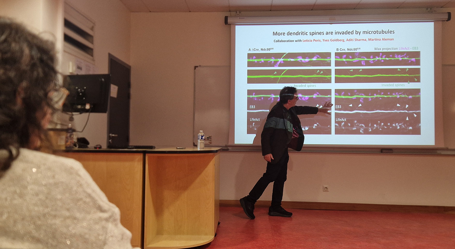
This observation finally helped connect the two phenomena: by entering dendritic spines more frequently, microtubules may promote their stabilization, thereby explaining the increase in spine number.
“This collaboration really was the missing piece that allowed us to connect what we knew about microtubules with the changes we were observing in neuronal morphology,” Schwarz emphasized.
Read also: The kinetochore: an unexpected player in neuronal connectivity
A New Mystery: Neuronal Survival
The researchers now had a coherent explanation for the role of kinetochore proteins in neuronal morphology.
“I could have happily retired at that point and said, ‘That's good, now we understand it!’” Schwarz joked.
But Guoli Zhao decided to take the investigation further.
Since removing NDC80 promoted axonal growth, Guoli Zhao wondered whether it might also enhance axon regeneration after injury. To test this hypothesis, he eliminated the protein from retinal ganglion cells in adult mice.
The result was striking: over the following weeks, large numbers of neurons disappeared. Additional experiments in cultured neurons confirmed that the loss of NDC80 triggered cell death through apoptosis.
“My reaction was, we're never going to figure out what's going on here,” Schwarz admitted.
How could a protein whose absence promoted neuronal process growth also be essential for neuronal survival?
To investigate this paradox, Guoli Zhao revisited an experiment that had helped establish the role of NDC80 in dendritic spine formation. He reintroduced a modified version of NDC80, unable to bind to microtubules, into neurons lacking the protein. And this time came another surprise: unlike what had been observed for dendritic spines, this modified version was sufficient to prevent neuronal death!
The researchers had found another clue: NDC80's roles in neuronal morphology and survival do not depend on the same molecular interactions.
But where do kinetochore proteins exert their protective function?
Further experiments, this time focusing on DSN1, another kinetochore protein, showed that a version restricted to the nucleus was sufficient to preserve neuronal survival.
These findings pointed towards a nuclear function for kinetochore proteins, possibly linked to the response to DNA damage.
Other observations supported this hypothesis: in the absence of DSN1, neurons accumulated higher levels of DNA damage markers. The researchers now suspect that certain kinetochore proteins may contribute to DNA repair, although this mechanism remains to be demonstrated.
New Avenues to Explore
What began with an unexpected mutation in fruit flies has revealed previously unsuspected functions for proteins once thought to be involved exclusively in cell division.
And the investigation is far from over.
Many questions remain unanswered. How are these proteins organized and regulated in neurons? Which other proteins do they interact with? And do they perform similar functions in other cells that no longer divide? The team is already exploring this last possibility in cardiac muscle cells.
With only a few years left before retirement, Thomas Schwarz now hopes that other researchers will pursue these lines of investigation. He humorously recalled something he had once been told: “When you're young, you're afraid that somebody will take your ideas. And when you're old, you're afraid that nobody will.”
Date
- Share
- Share on Facebook
- Share on LinkedIn